Developmental dysplasia of the hip
Developmental dysplasia of the hip is a serious pathology in neonates in which the femoral head is not in its normal position in the pelvic acetabulum. It represents a spectrum of pathologies, from slight dysplasia of the acetabulum, to complete dislocation of the femoral head. Developmental dysplasia of the hip is estimated to affect approximately one infant per thousand births.
-1vd17x.png)
Predisposing risk factors for developmental dysplasia of the hip include:
Pre-symptomatic ultrasound scan examination for developmental hip dysplasia is performed.
Our center offers pre-symptomatic screening for infants for early diagnosis of developmental hip dysplasia. Our aim is early diagnosis and treatment in order to reduce the risk of future surgical intervention.
Our recommendation for prevention, is to perform a clinical examination and a hip ultrasound scan in all infants with risk factors at the age of two weeks. In children without risk factors the test is not obligatory; our recommendation is to perform the test between the ages of 6-8 weeks following a referral by the paediatrician.
TREATMENT
It is important to note that treatment varies depending on the age at which the pathology is diagnosed. The sooner the diagnosis is made, the easier the treatment and the better the prognosis. Up to the age of six months, developmental hip dysplasia is usually treated conservatively with the application of a special Pavlik harness.
The image shows a baby treated with a Pavlik harness. The harness is flexible and aiming to keep the hips in flexion and abduction.

Failure of treatment or delayed diagnosis and presentation of the patient, leads to problems in gait and imbalance in childhood and pain and osteoarthritis in early adulthood.
If the diagnosis is delayed after the age of 6 months, surgical intervention is likely to be required. The type of surgical intervention depends on the degree of dislocation and the age of the child. The earlier the diagnosis is made, the better the long term outcome and the easier the procedure will be.
Specifically:
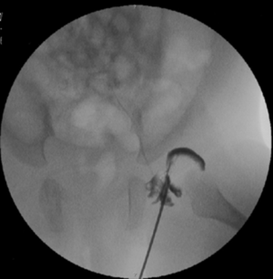
The x-ray shows a hip arthrography
performed by Dr. Zenios.
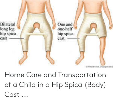
A child treated with a hip spica with hips in a reduced position

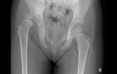
Pre-operative
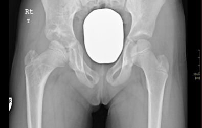
Post-operative
The x-rays show cases of developmental dysplasia of the in the hips that presented after the age of three years. The patients underwent surgery by Dr. Zenios for hip reduction with a pelvic and femoral osteotomy.
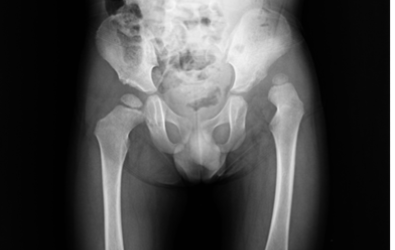
Pre-operative
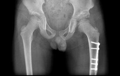
Post-operative